1. Before surgery Procedures
It all begins with an X-ray and CT scan of the knee joint that is further used to generate a 3D virtual model of the patient's anatomy. This virtual model is loaded into Mako software of LEO2 by Stryker Mako to create a personalized pre-operative plan.

X-ray
This helps the surgeon in seeing the damage initially before moving to the scanning stage.
X-rays are taken of AP standing, PA Flexion, Lateral and Sunrise position depending on case severity.
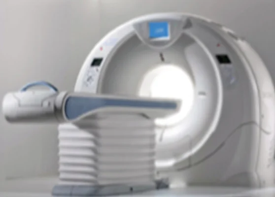
CT-scan
A motion rod is placed near the patient for accuracy during the scanning process.

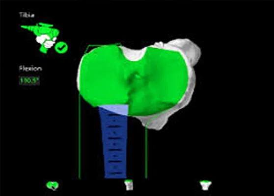
Segmentation
The CT scan data is loaded and converted into the Mako software.
Post this, landmarks are identified and a virtual model of the bone in 3d and planning images are created.
2. LEO2 by Stryker Mako In the Operation Theater
In the operation theatre, the surgeon uses the LEO2 by Stryker Mako system to prepare the bone for the implant and guides the LEO2 robotic arm within the predefined area and stays within the planned boundaries.
3. Post surgery
The AIOR team of surgeons, nurses and physical therapists will monitor condition and progress and review the post-operative x-ray of the implant.
